Western Kentucky Diagnostic Imaging A Department of The Medical Center
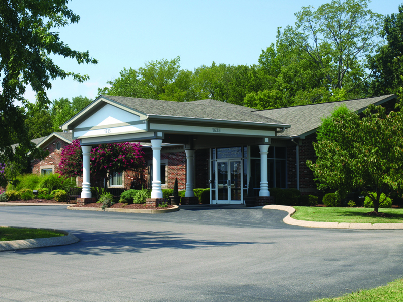
- 1635 Scottsville RoadBowling Green, KY 42104Get Directions
Hours of Operation
Monday - Friday
- 8:00 am - 5:00 pm
- Scheduling: (270) 745-1199
- Phone: (270) 780-2720
- Toll Free: (888) 746-9500
- Fax: (270) 746-9113
Western Kentucky Diagnostic Imaging, a department of The Medical Center, offers exceptional quality in clinical service, images and diagnoses, which is critical to determining your care and treatment. Our facility has a caring team committed to providing outstanding service for all patients and referring physicians. A comfortable and relaxed environment is assured when you choose Western Kentucky Diagnostic Imaging for imaging services!
Western Kentucky Diagnostic Imaging (WKDI), a department of The Medical Center, has been designated a Breast Imaging Center of Excellence by the American College of Radiology (ACR). WKDI is the first facility in Southcentral Kentucky to receive this designation. By awarding facilities the status of Breast Imaging Center of Excellence, the ACR recognizes breast imaging centers that have earned accreditation in mammography, stereotactic breast biopsy, breast MRI and breast ultrasound (including ultrasound-guided breast biopsy).


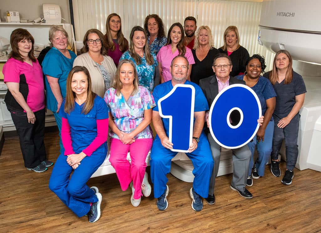
WKDI Celebrates 10th Anniversary
Western Kentucky Diagnostic Imaging (WKDI) celebrated its 10th anniversary on September 30, honoring the facility’s decade of service to the Bowling Green community.
WKDI offers exceptional quality in clinical service, images, and diagnoses, which is critical to determining your care and proper treatment.
Med Center Health purchased WKDI in October of 2012, and the Director of Radiologic Services, Eddie Scott, spoke about the changes and improvements the facility has seen over the last decade.
“We’re continuing to invest in new technology,” said Scott. “Since inception 10 years ago, we have purchased and installed a new open MRI scanner. We’re very proud of that. A new CT scanner has been purchased, as well as new X-ray and ultrasound equipment. We’re continually looking to invest in new technology to keep WKDI on the cutting edge, and we’re proud that we’re able to do that.”
WKDI was originally established in 1995, and nearly half of the technical and clerical staff currently working at WKDI have been at the facility for more than 10 years.
“Brian Stephens, the manager here on site, has done a tremendous job, and he has been with WKDI since 2000, some 12 years prior to our purchase of the facility,” said Scott. “We’re proud of WKDI. They do a tremendous job, and they certainly have embraced the goal of all of our imaging sections at Med Center Health of providing outstanding care to all of our patients in a very loving, caring, effective, efficient and safe manner with everything we do revolving around the care of our patient and the patient’s family.”
Western Kentucky Diagnostic Imaging (WKDI), a department of The Medical Center, has been designated a Breast Imaging Center of Excellence by the American College of Radiology (ACR). WKDI is the first facility in Southcentral Kentucky to receive this designation.
Patient Services
- Low-dose CT Scans for Lung Cancer
- High Field Open MRI
- 3D Digital Mammography
- Stereotactic Breast Biopsy
- Ultrasound
- Bone Density Evaluation
- Fluoroscopy
- CT Scan
- X-Ray
- Vertebral Fracture Assessment
-
1635 Scottsville RoadBowling Green, KY 42104